

ODM Thoracic Aortic Stent Graft System for Minimally Invasive Aortic Aneurysm Repair
2026-10-12
What if repairing a thoracic aortic aneurysm no longer demanded cracking open the chest? With the ODM Thoracic Aortic Stent Graft System, INT is turning that possibility into a minimally invasive reality—one that could redefine recovery times and patient outcomes.
Turning a Major Aortic Repair into a Small Groin Puncture
Repairing the aorta once meant opening the chest or abdomen, but many of those extensive procedures now begin and end with nothing more than a small puncture in the groin. Catheters and stent grafts are advanced through the femoral artery while the surgical team watches on live imaging. The aorta is relined from within, and the only external mark is a nick that can be covered with a bandage.
This shift changes the entire recovery timeline. Instead of days in intensive care after a large incision, patients are often sitting up within hours and walking the next morning. Closure devices seal the puncture site from inside the vessel, so there’s no need for prolonged pressure or a second incision. For older or frail patients who would struggle with open surgery, this approach turns a major aortic repair into a manageable, same-day access procedure.
The Small Design Decisions That Prevent Endoleaks
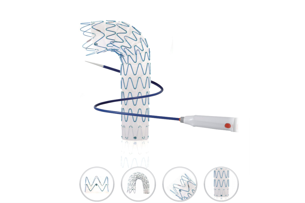
Endoleaks rarely stem from a single catastrophic oversight. More often, they trace back to a series of tiny, seemingly inconsequential choices made during stent-graft sizing, overlap planning, or seal zone preparation. The difference between a durable seal and a late type Ia leak can hinge on a millimeter of oversizing, the angle of a proximal landing zone, or how aggressively you treat a calcified neck before deployment.
Consider the proximal seal as a negotiation between the graft fabric and the aortic wall. Too little radial force, and the fabric loses contact during cardiac pulsation. Too much, and you invite intimal damage that evolves into a new channel over time. Skilled operators learn to read the pre-procedure CT as a dynamic structure, not a static blueprint, adjusting graft diameter not to a fixed percentage but to the local compliance, thrombus burden, and curvature of each specific neck.
Small decisions also govern the appendages: choosing a flared limb for a conical iliac, extending into the external iliac when the internal is diseased, or adding a proximal cuff solely to lengthen a marginal neck by a few critical millimeters. These steps rarely feel heroic at the time, but across hundreds of cases they separate the repairs that remain silent from those that return with a faint, delayed blush on surveillance imaging.
When a Thoracic Stent Graft Makes More Sense Than Open Surgery
For many patients with descending thoracic aortic aneurysms or complicated type B dissections, the decision between a stent graft and open repair hinges on physiology more than anatomy. The minimally invasive option spares the patient a thoracotomy, single-lung ventilation, and the profound inflammatory cascade that follows aortic cross-clamping. Frail individuals, those with severe COPD, or anyone who has already weathered a prior chest operation often cannot afford the prolonged recovery and high perioperative risk of open surgery. A stent graft, delivered through a small groin incision, shifts the calculus dramatically: shorter ICU stays, less blood loss, and a faster return to baseline function.
Anatomy still matters, but the envelope for endovascular repair has expanded. Favorable landing zones, adequate iliac access, and the absence of critical branch vessel involvement make a thoracic stent graft not just feasible but preferable. When the pathology is confined to the straight segment of the descending aorta and the seal zones are healthy, the device can exclude the aneurysm or cover the primary entry tear of a dissection without the need for cardiopulmonary bypass. This translates into lower rates of paraplegia when compared to open repair in matched cohorts, particularly if spinal cord protective measures like lumbar drains and staged coverage are employed.
There are also economic and logistical arguments that tip the scale. Hospitals without a dedicated aortic surgery team can often perform an endovascular procedure with interventional radiology and vascular surgery collaboration. The learning curve is shorter, the operative time is more predictable, and the demand on blood bank resources is minimal. For the patient, the difference is visceral: a three-day hospital stay versus three weeks, a small scar versus a long posterolateral incision, and the ability to resume normal activities in weeks rather than months. When the clinical picture is borderline, the stent graft offers a less destructive path that preserves future options, whereas open surgery burns bridges and leaves a lasting physiologic debt.
How the ODM System Handles Challenging Aortic Arch Anatomy
The ODM system addresses aortic arch challenges by allowing incremental adjustment during deployment. Unlike rigid delivery systems, its multi-stage release mechanism lets the operator fine-tune the proximal sealing zone even in steeply angled arches, where a type III configuration leaves minimal room for error. The system's torque-responsive handle translates subtle rotational inputs into controlled advancement of the stent graft, reducing the tendency for the device to “watermelon seed” forward when crossing the arch.
Another key feature is the low-profile, pre-shaped tip that tracks through tortuous iliac and arch segments without straightening the native anatomy. For gothic arches with acute takeoff angles, the tip's graduated stiffness transition prevents excessive force at the inner curvature, protecting the fragile intima from dissection. Meanwhile, the proximal clasp mechanism maintains partial constraint until the operator confirms orientation relative to the greater curvature, allowing rotational correction without losing position.
Post-deployment, the ODM system's optional balloon-expandable sealing ring can be inflated at a lower pressure to accommodate irregular landing zones, such as those with focal calcification or a retro-esophageal course. This adaptive strategy reduces bird-beak configuration and endoleak risk in angulated arches, where conventional cylindrical devices often fail to appose the lesser curvature.
A Hospital Stay Measured in Days Instead of Weeks
Not long ago, a routine surgery meant packing a bag for a week. Now, thanks to laparoscopic tools, targeted anesthesia, and aggressive early mobility protocols, many patients are walking out the door in under 72 hours. The shift isn't about rushing people home—it's about removing the reasons they had to stay so long in the first place.
Postoperative pain used to be managed with heavy narcotics that kept patients groggy and bedridden. Today, regional nerve blocks and multimodal analgesia let people breathe deeply, eat sooner, and move without the fog. That simple change collapses recovery timelines, cutting the average stay from fourteen days to three or four in many orthopedic and abdominal cases.
Even complex procedures like joint replacements are being reclassified as outpatient or short-stay surgeries. Hospitals now design dedicated fast-track units where physical therapy starts hours after the operation, not days later. The result is fewer hospital-acquired infections, less disruption to family life, and a bill that doesn't spiral out of control.
What Procedure Logs Reveal About ODM Graft Performance
Surgical logs offer an unfiltered view of how ODM grafts behave outside controlled trials. The notes capture intraoperative handling, initial fixation stability, and early postoperative reactions—details that often go unreported in formal studies. A recurring pattern is the graft's consistent shape retention during placement, which reduces operative time and minimizes the need for trimming, but logs also flag instances of minor particulate migration when the recipient bed is poorly prepared.
Longitudinal entries from follow-up visits reveal the graft's integration timeline more reliably than retrospective surveys. Clinicians record bone density changes at the interface, soft tissue response, and any signs of delayed union. In several logs, patients with compromised vascularity showed slower incorporation, while those with healthy cancellous beds demonstrated predictable remodeling within expected windows. The handwritten or electronic notes also capture rare events like early resorption or low-grade inflammation, providing a granular risk profile.
Aggregated procedure logs expose performance variability across patient subgroups that randomized data often misses. Smokers, diabetics, and patients on long-term corticosteroids appear to have extended healing phases, yet the graft still maintains structural integrity in most cases when load-bearing is delayed appropriately. These real-world observations help refine preoperative planning and postoperative protocols, turning routine documentation into a practical feedback loop for clinicians.
FAQ
It uses a low-profile delivery system with a multi-segmented nitinol frame, which improves conformability in angulated aortic arches while maintaining radial force at the proximal and distal landing zones.
Patients with descending thoracic aortic aneurysms, penetrating ulcers, or traumatic aortic transections who have suitable vascular access and proximal and distal seal zones are generally candidates after imaging review.
Because it is delivered through a small femoral or iliac access site, most patients experience shorter hospital stays, less blood loss, and faster return to normal activity than with open thoracotomy.
It is offered in a broad range of diameters and lengths, with tapered and flared configurations available to accommodate differing aortic neck and iliac anatomies.
When the proximal landing zone is adequate or when used with arch debranching or chimney techniques, it can be deployed in selected arch cases, though this depends on the operator's judgment and patient-specific anatomy.
Contrast-enhanced CT angiography is typically performed at one, six, and twelve months, then annually, to monitor for endoleak, migration, or aneurysm sac expansion.
The low-porosity polyester or ePTFE fabric is tightly woven and attached to the stent frame with minimal suture holes, which helps limit transmural flow and encourages early sac thrombosis.
It features a hydrophilic-coated introducer sheath with sizes ranging from 18F to 24F, allowing navigation through tortuous iliofemoral vessels while preserving controlled deployment.
Conclusion
The ODM Thoracic Aortic Stent Graft System turns what used to be a major open chest procedure into a small groin puncture, with the graft delivered through the femoral artery under fluoroscopic guidance. This minimally invasive route avoids sternotomy, cardiopulmonary bypass, and aortic cross-clamping, which means a hospital stay measured in days rather than weeks. The device’s design focuses heavily on seal integrity: the proximal and distal anchoring zones, fabric weave, and radial force of the stent skeleton are engineered to prevent type I and type III endoleaks, which remain the main cause of late aneurysm sac pressurization. These are deliberate, small design decisions—not just material tweaks—that keep blood from re-entering the sac. For challenging aortic arch anatomy with acute angulation, short landing zones, or heavy calcification, the ODM graft uses a flexible delivery system and controlled deployment to maintain wall apposition without sacrificing conformability.
Procedure logs from real-world use show consistent technical success and low reintervention rates across varied patient anatomies. In many cases, the ODM system makes more sense than open surgery, especially for older patients or those with significant comorbidities who would struggle with a thoracotomy. The combination of percutaneous femoral access, tailored sealing zones, and adaptable arch conformability reduces physiological stress while preserving durable aneurysm exclusion. Rather than relying on broad claims, the accumulating case data reflect the practical impact: shorter ventilation times, fewer ICU days, faster discharge, and fewer access-site complications. This shift from open repair to a groin puncture with a thoracic stent graft is not just about smaller incisions—it changes the risk calculus for patients and surgeons alike, offering a repair option that matches the anatomy while drastically cutting recovery burden.
Contact Us
Contact Person: Jeffrey
Email: [email protected]
Tel/WhatsApp: 86-0633-2230056
Website: https://www.sd-intmedical.com